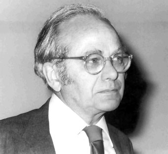
David Hunter Hubel
13 February 2026
David Hunter Hubel
(Windsor, 1926 – Lincoln, 2013) Canadian-born American neurobiologist. He studied medicine at McGill University in Montreal, where he received his doctorate in 1951, and worked in Montreal and Baltimore. He became a naturalized U.S. citizen in 1953, and from 1959 he taught at Harvard University, where he chaired the Department of Neurophysiology from 1967 to 1982, the year he moved to John Franklin University.
 Together with his colleague Torsten Wiesel, David H. Hubel embarked in 1958 on a series of investigations aimed at understanding the mechanism of visual perception at the cortical level. They used microelectrodes and modern chemical techniques to detect and analyze the activity of neurons in the brain region called the visual cortex (specifically, area 17), composed of millions of neurons responsible for processing images received through the retina and optic nerve.
Hubel and Wiesel discovered that the activity of nerve cells in the cortex in response to visual stimulation is highly specific. Some cells respond only to patches of light, while others respond to a line whose inclination is critical, such that the response can be altered by a change in angle of just ten degrees. They also described the organization of these specific cells into columns, classifiable into various types according to their function. Thanks to their contributions, the visual cortex is currently the best-understood part of the brain. David Hubel and Torsten Wiesel were awarded the Nobel Prize in Medicine in 1981 (shared with American neurobiologist Roger Wolcott Sperry) for their discoveries concerning information processing in the visual cortex.
The passing of a scientific figure who revolutionized our understanding of the brain is a cultural and emotional loss for those of us who knew him. This is the case of Professor David H. Hubel, who died on September 22, 2013, in Lincoln, Massachusetts, at the age of 87. His wife, Ruth, had passed away the previous February.
Professors David Hubel and Torsten Wiesel received the Nobel Prize in Physiology or Medicine in 1981. This award recognized their most significant contributions to the understanding of the physiology of the visual system, the functional organization of the brain, and the multidisciplinary approach to its study. Since then, Professor Hubel continued to make fundamental contributions to the understanding of the visual system, contributing significantly to the intellectual revolution in neuroscience that we have witnessed in recent years.
To fully grasp the significance of their contributions, we must remember that the concepts and knowledge of brain physiology, and of the visual system in particular, in the 1950s, when Hubel and Wiesel began their work at Johns Hopkins Medical School, were different and very limited compared to today. At that time, the question of brain localization was being debated, and electrophysiology relied primarily on electroencephalographic recordings, evoked potentials, and electrical stimulation. The introduction of the microelectrode as a technique for recording the activity of a neuron—the functional unit of the brain—represented a significant qualitative shift, as it allowed scientists to understand the responses of these cells to natural stimuli. Professor Hubel also strove to prevent the separation of the components of what we now call Neuroscience—anatomy, physiology, and biochemistry—and to unite them.
David Hunter Hubel was born in Windsor, Ontario, Canada on February 27, 1926. He studied medicine at McGill University and, in 1955, became a Research Fellow at the Walter Reed Army Institute of Research in the USA. From there, he moved to Johns Hopkins Medical School in Baltimore, and a year later to Harvard Medical School, where he has continued his work, demonstrating a clear loyalty to the institution. Professor Hubel’s contributions to the understanding of visual physiology have been fundamental and can be summarized in four main achievements.
David Hubel, along with Torsten Wiesel, demonstrated that cells in the visual cortex are capable of detecting appropriate visual images; that is, visual cortex cells are selective for stimulus patterns. They showed that the most suitable stimulus pattern is not the same along the visual pathway, from the retina to the visual cortex, and that the stimulus to which visual cells respond is one formed by contrasting edges with a specific orientation. The implications of these discoveries were revolutionary. The visual cortex was no longer thought of as a structure of thousands of cells, each participating in the reconstruction of the visual scene. The change implies that each cell is activated only by its own stimulus, meaning that when it is activated, it is signaling something specific about the nature of the image in a particular region of the visual field.
In the visual cortex, they discovered neurons that are activated when a stimulus enters through either eye and that the receptive field of each eye was located approximately in the same region of the visual field. This implies that when the eyes are aligned, an object in the visual scene will simultaneously activate the same visual cell through both eyes. The discovery of these binocular cells is particularly important for explaining depth perception, or stereopsis, since comparing the images from both eyes allows the brain to determine the relative position of objects in space.
In the visual cortex, there is a detailed representation of the visual field. Hubel and Wiesel’s third major contribution was revealing the functional microstructure of the visual cortex. In each mm³ of the visual cortex, they discovered a regular distribution of ocular dominance—that is, cells that prefer to be activated by one eye or the other—and orientation axes—cells that prefer a stimulus pattern with a specific orientation. Rafael Lorente de Nó, in 1938, had already described an anatomical organization of neurons in the cerebral cortex in columns, and in the 1960s, Vernon B. Mountcastle, in the somatosensory cortex, had already discovered that the columns of cells shared the same functional characteristics. From there, Hubel and Wiesel discovered that in the visual cortex, neurons are arranged according to their preference for the orientation of stimuli and the eye through which those stimuli arrive.
The fourth important contribution, with significant clinical implications, has been understanding the temporal development of the plasticity of the visual system. A strabismic eye ends up performing its function much worse than a healthy one. Hubel and Wiesel defined the critical age range within which the development of the visual cortex is modified by experience, the ontogenetic determinism of the function of the visual system, but also the plasticity of the response to changes in visual experience. These findings have had a major impact on ophthalmological practice.
Both David Hubel and Torsten Wiesel have been closely associated with the Neuroscience Laboratories at the University of Santiago de Compostela. It all began with a research program on the visual system that led postdoctoral fellows from my laboratory to continue their training in the neuroscience of vision outside of Spain. The first was Dr. Francisco González, who completed a postdoctoral fellowship with Dr. Gian F. Poggio, the discoverer of neurons in the visual cortex that respond to retinal disparity, at the Department of Physiology of Johns Hopkins University in 1984. Then, in 1990, Dr. Javier Cudeiro moved to the Institute of Ophthalmology at University College London to work with Dr. Adam Sillito, who, combining pharmacological and electrophysiological techniques, had demonstrated the thalamo-cortico-cortical interrelationships in visual function, research that Dr. Cudeiro would later continue at the University of A Coruña. Next was Dr. José Manuel Alonso, who, after an interview with Torsten Wiesel at a conference in Alicante in 1992, continued his work at Rockefeller University and is currently at the State University of New York.